All the examples shown below were obtained using this variant: TP53:p.R175L.
How to access the protein viewer tool in VarSome Clinical
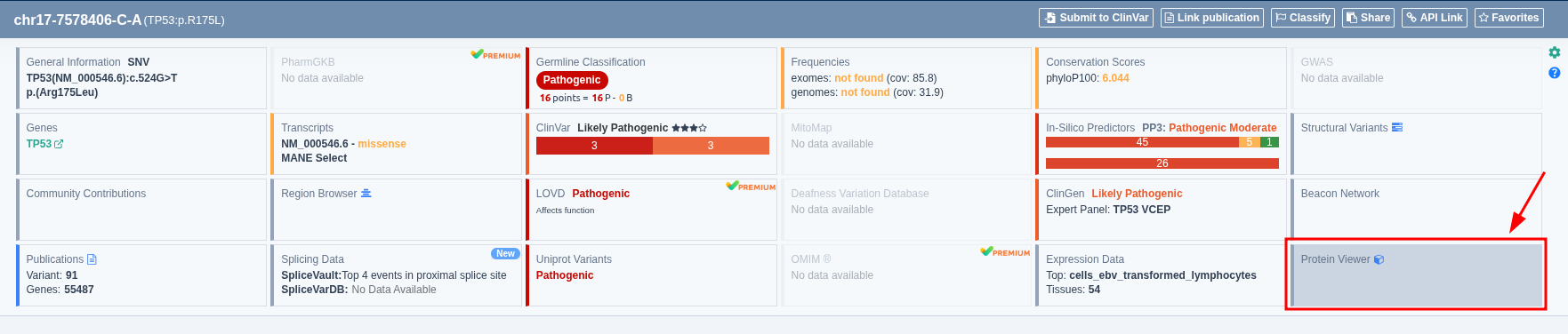
This tool is accessible from the “Gene/Variant basic information” section. Click on the “Protein Viewer” button to open a new window with the 3D Protein Viewer tool.
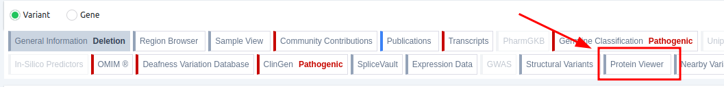
From the Variant table
From the Gene table

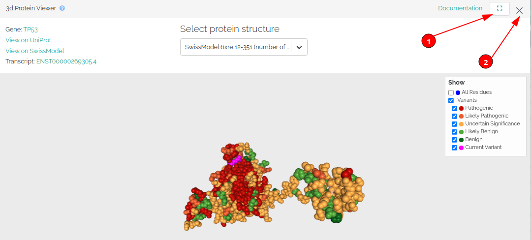
Let’s use the variant TP53:R175L as an example on the 3D Protein Viewer.
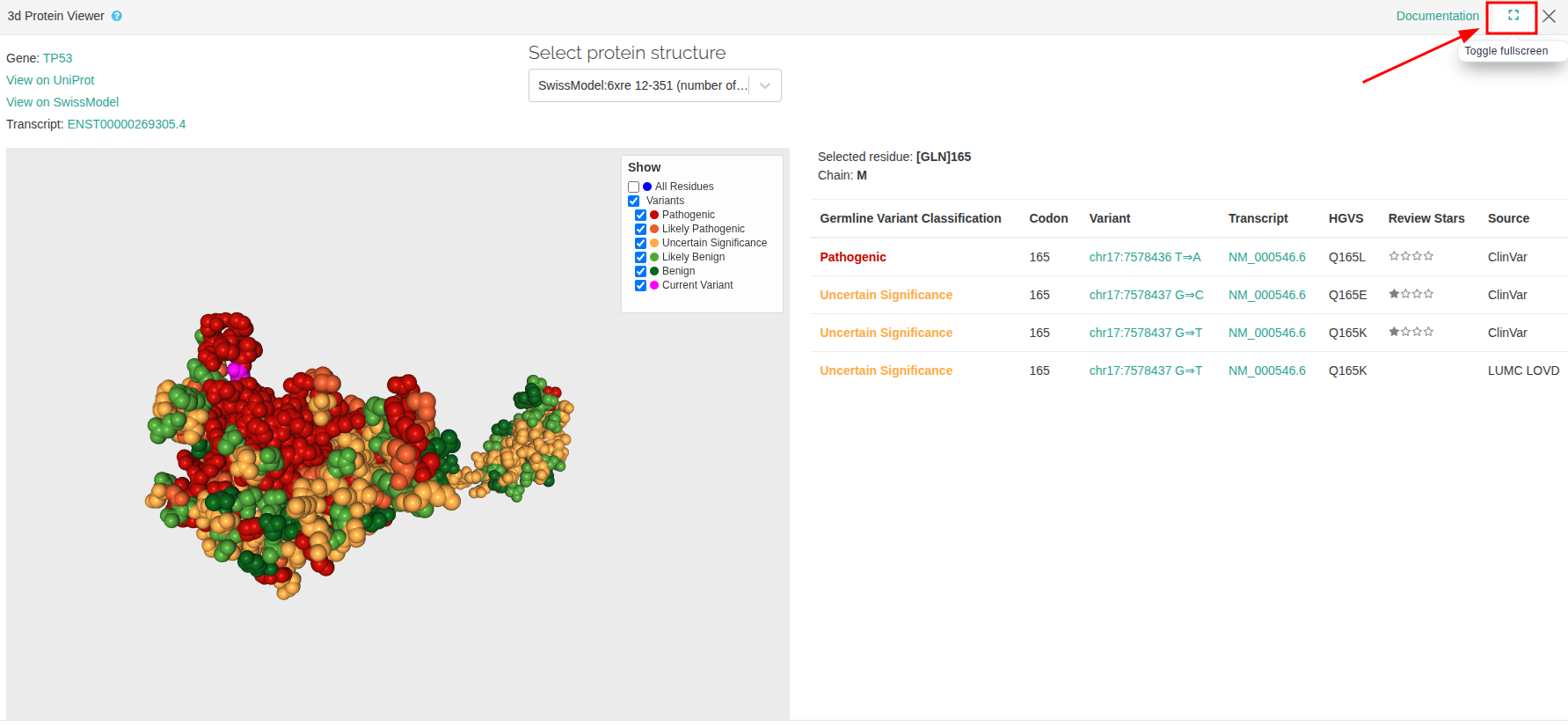
Go to the top right corner of the window to maximize the protein viewer page (1) or to close the window (2) and keep navigating through the VarSome website.
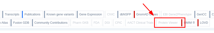
Please note that if there is no available structure, the "Protein Viewer" icon will be grayed out:
How to display variants on the protein structure
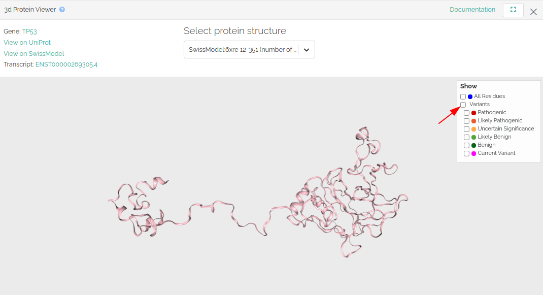
Variants reported from ClinVar, Uniprot and the VarSome Community are mapped onto the protein structure and are colored according to the Germline Variant Classification. Additionally, you will be able to see the variant of interest (the variant whose VarSome page you are currently visiting) in pink. You can decide which variants are shown in the "Show" menu on the top right corner.
- Click on “Variants” to deselect all variants and display the protein structure without highlighting any residue position.
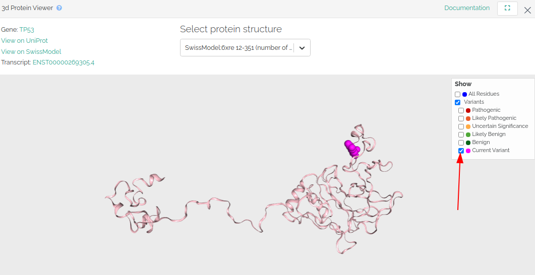
- Click on "current variant" to show the variant you have searched.
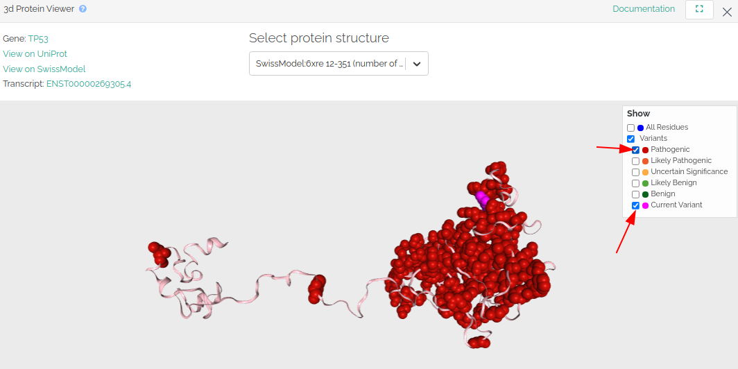
- Click on the other variant options to start adding variants to the protein structure. For example, you can select "current variant" and "pathogenic" variants to check whether your variant clusters with the reported pathogenic variants in that gene.
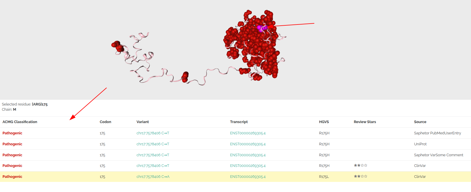
- Click on a residue of the protein structure to show a list of reported variants at that position below the protein structure.
How to use the protein viewer camera
- Use the left mouse button to rotate the view.

- Use the right mouse button to pan (move the camera horizontally).

- Use the mouse wheel to zoom in / out.

- Double-click with the left mouse button to focus.

This feature enables when entering the full-screen mode to see all the related variants on the right-hand side:
How to select a different protein structure
We import protein structures from Swiss-Model. The protein structure shown by default will be:
- The structure that contains the variant of interest, if available.
- The structure with the highest number of pathogenic variants.
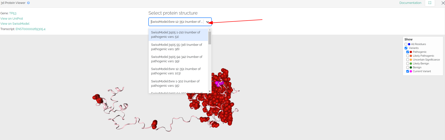
Click the arrow next to the protein structure's name to select a different protein structure from the drop-down menu.
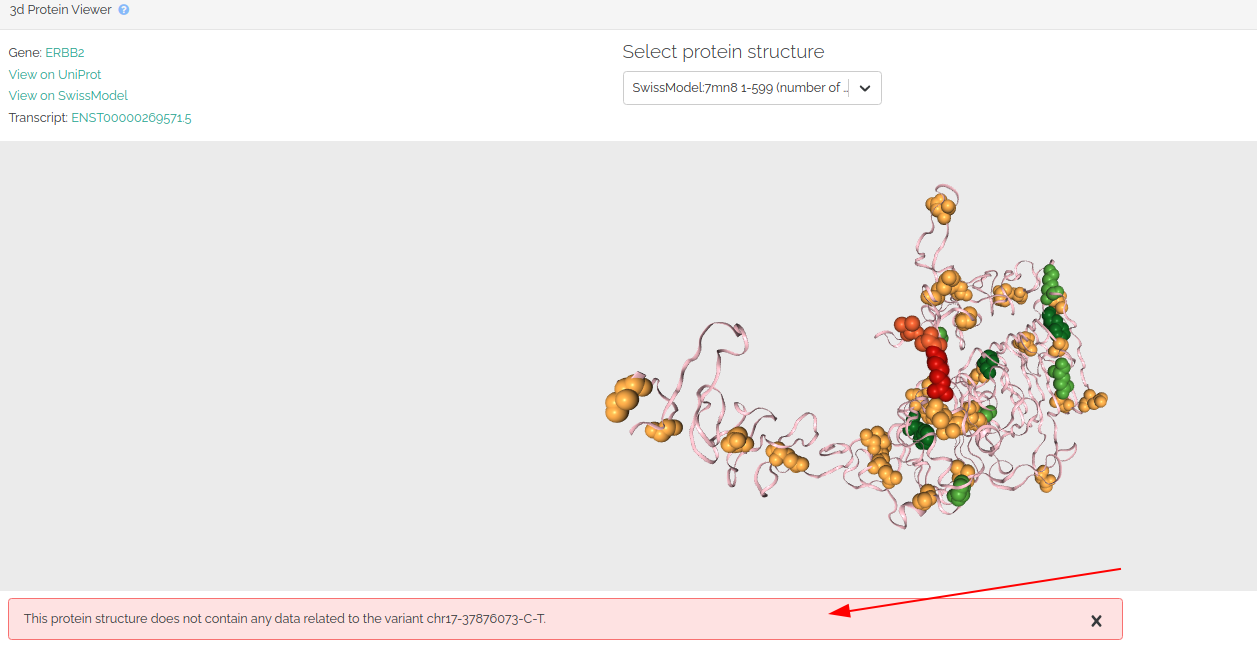
Sometimes the selected protein structure does not contain any data related to the variant you have searched. If this is the case, a message below the structure will be shown to notify the user.
If you experience issues when using the 3D protein, please try to clearing your browser data (e.g. cookies) before continue navigating. Please, contact us if the issue persists.
How to access the protein viewer tool in VarSome
The Protein Viewer tool can be accessed by clicking the card "Protein Viewer" as shown below: